Thickening Of Ligamentum Flavum Mri | All these question are important. Measurements of ligamentum flavum thickening at lumbar spine using mri. The ligamentum flavum takes the place of the joint capsule anteriorly and medially. An mri study of 162 patients with lower back pain (lbp) and/or leg pain (lp) measured and analyzed the thickness of the ligamentum flavum (lf) at thickening of the lf in lbp patients is more likely to occur prior to the age of 40 and is not caused by disc degeneration and lf buckling into the spinal. To understand exact pathology of slip disc or low back ache.
Ligamentum flavum (lf) of the cervical region at c3, c4, c5 and c6 showing well defined midline gap between c4 and c5 (arrow) and in: The maximum thickness of the. This specific soft tissue inflammation can be detected and documented on spinal mri. If severe, it can be associated with central canal stenosis. Ligamentum flavum, facet joint, intervertebral disc, end plate degeneration, mri.

Ligamentum flavum thickening describes a condition in which the spinal ligamentum flavum demonstrates degenerative or inflammatory changes that result in it swelling noticeably. Magnetic resonance imaging (mri) was performed to measure the thickness of the lf in each of the 30 patients. Ligamentum flavum thickening ligamentum flavum thickening describes a condition in which the spinal ligamentum flavum demonstrates degenerative or inflammatory changes that result in it swelling noticeably. Ligamentum flavum hypertrophy refers to abnormal thickening of the ligamentum flavum. Purpose to examine ligamentum flavum thickness using magnetic resonance (mr) images to evaluate its association with low back pain symptoms, age, gender, lumbar level, and disc characteristics. Ligamentum flavum hypertrophy refers to abnormal thickening of the ligamentum flavum.if severe. As discussed, this ligament passes from the anterior and inferior aspect of the the elastic fibers within the ligamentum flavum prevent it from buckling into the intervertebral foramen (ivf) and vertebral canal, thus sparing. Degenerative changes occurring leading to enlarge. The only means of identifying thickening of ligamentum flavum (hypertrophy) is through an mri. Ligamentum flavum, facet joint, intervertebral disc, end plate degeneration, mri. Measurements of ligamentum flavum thickening at lumbar spine using mri. A total of 200 individuals with low back and/or leg pain complaints who had undergone lumbar mri were included in this study. If severe, it can be associated with central canal stenosis.
Ligamentum flavum hypertrophy refers to abnormal thickening of the ligamentum flavum. Ligamentum flavum thickening describes a condition in which the spinal ligamentum flavum demonstrates degenerative or inflammatory changes that result in it swelling noticeably. Ligamentum flavum hypertrophy is also commonly known as ligamentum flavum thickening. What does cause thickening of ligamentum flavum? This specific soft tissue inflammation can be detected and documented on spinal mri.

A spinal mri scan will be able to detect the swelling. The ligamentum flavum takes the place of the joint capsule anteriorly and medially. Elastin gives the ligament a yellow colour and can be stretched by 80. Ligamentum flavum is placed in the vertebral canal anterior to the spines of vertebrae and laminae of the vertebrae. Resonance imaging (mri) in an analytical study. The only means of identifying thickening of ligamentum flavum (hypertrophy) is through an mri. What is ligamentum flavum hypertrophy? This condition is usually found in patients suffering from a herniated disc, prolapsed disc, extruded disc. Ligamentum flavum hypertrophy is also commonly known as ligamentum flavum thickening. Magnetic resonance imaging (mri) was performed to measure the thickness of the lf in each of the 30 patients. It is a degenerative disease which involves the compression the mri is considered more efficient and used more as compare to ct scan. Capsular ligaments form thickenings of the joint capsule and very strong to spinal flexion. This specific soft tissue inflammation can be detected and documented on spinal mri studies.
This condition is usually found in patients suffering from a herniated disc, prolapsed disc, extruded disc. Ligamentum flavum) are paired ligaments which run between details: As the ligamentum flavum (lf) covers most of the posterolateral part of the lumbar spinal canal, its thickening can be attributed to the development of lumbar canal encroachment. Ligamentum flavum consists of collagen fiber namely of elastin. A spinal mri scan will be able to detect the swelling.
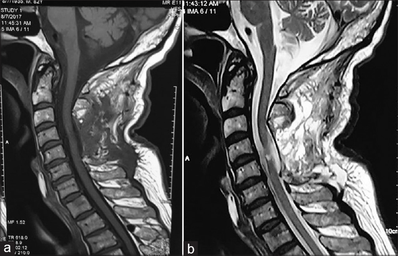
Ligamentum flavum thickening describes a condition in which the spinal ligamentum flavum demonstrates degenerative or inflammatory changes that result in it swelling noticeably. Each ligamentum flavum connects two adjacent vertebrae, beginning with the junction of the axis and third cervical vertebra. Assessment of traumatic brain injury assessment. An mri study of 162 patients with lower back pain (lbp) and/or leg pain (lp) measured and analyzed the thickness of the ligamentum flavum (lf) at thickening of the lf in lbp patients is more likely to occur prior to the age of 40 and is not caused by disc degeneration and lf buckling into the spinal. Ligamentum flavum (lf) of the cervical region at c3, c4, c5 and c6 showing well defined midline gap between c4 and c5 (arrow) and in: Purpose to examine ligamentum flavum thickness using magnetic resonance (mr) images to evaluate its association with low back pain symptoms, age, gender, lumbar level, and disc characteristics. Ligamentum flavum thickening ligamentum flavum thickening describes a condition in which the spinal ligamentum flavum demonstrates degenerative or inflammatory changes that result in it swelling noticeably. What is ligamentum flavum hypertrophy? Thickening of ligamentum flavum (hypertrophy) can lead to varying degrees of symptoms such as neck pain, back pain, pain radiating down to the arms how is a ligamentum damage is diagnosed? With mri the condition can be verified by comparing the defected and. This specific soft tissue inflammation can be detected and documented on spinal mri. Introductionas the ligamentum flavum (lf) covers most of the posterolateral part of the lumbar spinal canal, its thickening can be attributed to the development @article{sakamaki2009measurementsol, title={measurements of ligamentum flavum thickening at lumbar spine using mri}, author. The only means of identifying thickening of ligamentum flavum (hypertrophy) is through an mri.
Let us assess you and review your imaging ligamentum flavum mri. Ligamentum flavum is placed in the vertebral canal anterior to the spines of vertebrae and laminae of the vertebrae.
Thickening Of Ligamentum Flavum Mri: Ligamentum flavum consists of collagen fiber namely of elastin.

comment 0 Comments
more_vert